Custom Recombinant Lentivirus Service
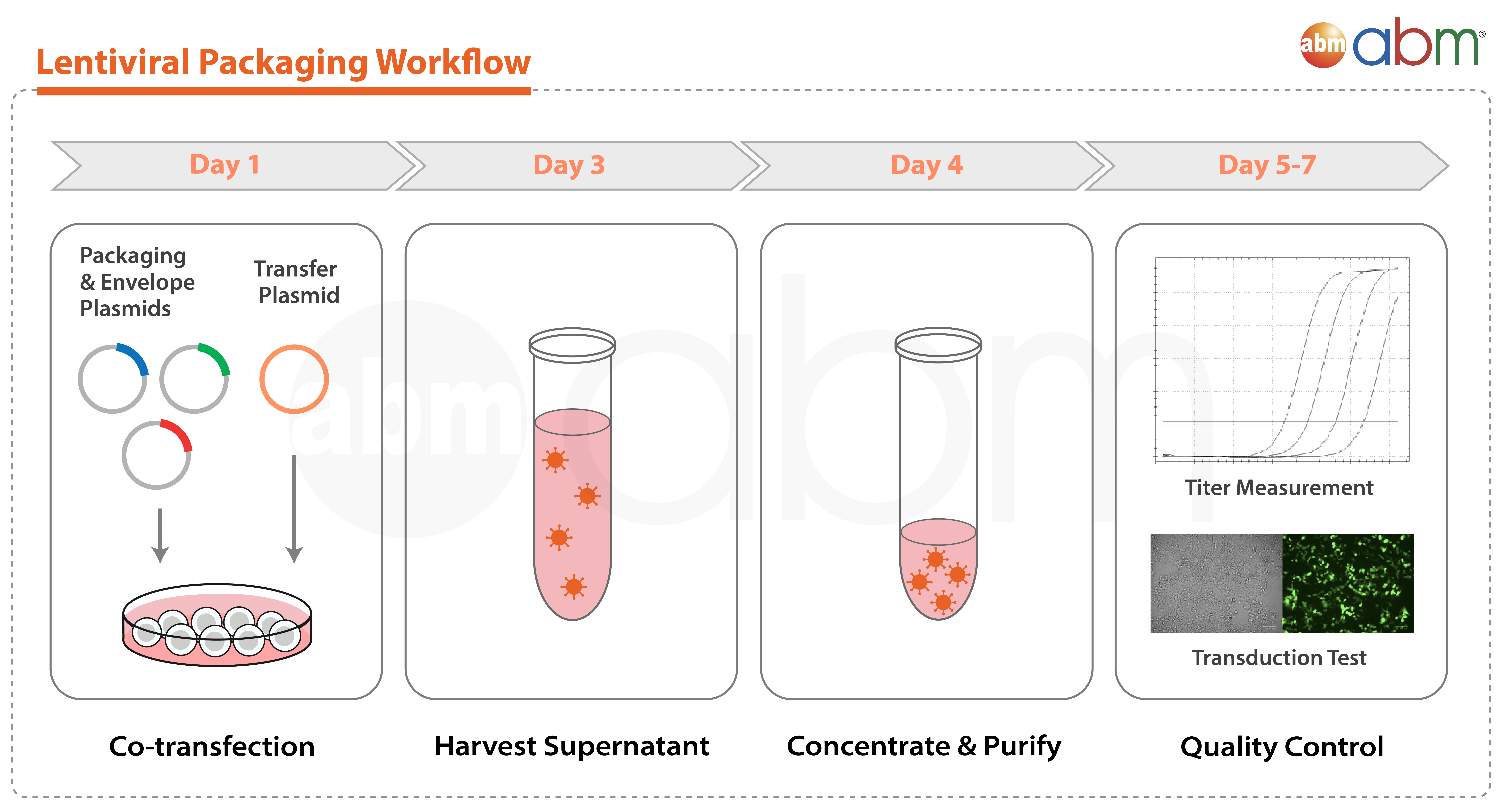
As a leading expert in lentiviral technology, abm provides a Custom Lentivirus Service to support the design, cloning, and viral packaging of your unique lentivirus construct. We offer a range of research-grade lentivirus with ultra-high titers up to 10¹⁰ IU/ml to meet your experimental requirements. Need something different? Explore our ready-to-use gene libraries or inquire with us below to get started on your Custom Lentivirus project.
Advantages of the Lentivirus System:
- High infection efficiency
- Stable integration & expression of recombinant proteins
- Large insert capacity (up to 5kb)
- Ideal system for the creation of stable cell lines
- Broad host range: infection of dividing, non-dividing, stem cells, and primary cells
Service Details
Gene Synthesis & Cloning Services
| Service | Unit | Cat. No. | Price |
|---|---|---|---|
| Gene Synthesis | Per bp | C098 | Starting at USD $0.18/bp |
| Subcloning Service I | 1.0 μg | C096 | $125.00 |
| Subcloning Service II | 1.0 μg | C097 | $225.00 |
Custom Lentivirus Packaging Services
| Service | Unit | Titer | Purity | Application | Cat. No. | Lead Time | Price |
|---|---|---|---|---|---|---|---|
| Standard Titer Custom Recombinant Lentivirus Packaging | 4 x 500 μl | 107 IU/ml | Research-grade | Standard cell culture | LV001‑a | 2-3 weeks | $199 |
| Medium Titer Custom Recombinant Lentivirus Packaging | 4 x 250 μl | 108 IU/ml | Research-grade | Standard cell culture | LV001‑b | 2-3 weeks | $449 |
| High Titer Custom Recombinant Lentivirus Packaging | 10 x 100 μl | 109 IU/ml | Research-grade | Difficult to transduce cells | LV001‑c | 2-3 weeks | $749 |
| Ultra-High Titer Custom Recombinant Lentivirus Packaging | 10 x 100 μl | 1010 IU/ml | Research-grade | Difficult to transduce cells | LV001‑d | 2-3 weeks | $1049 |
Add-on Services
| Service | Unit | Cat. No. | Price |
|---|---|---|---|
| Custom Lentivirus Aliquoting | Up to 10 Vials | C507 | $50.00 |
| Custom Lentivirus Titration - qPCR-based (Detectable Range of 104-1010 IU/ml) | 1 Sample | C099 | $85 |
| Plasmid Amplification for Virus Packaging Service | 1 Service | C314 | $100.00 |
Custom Pseudotype Lentivirus
| Service | Unit | Cat. No. | Price |
|---|---|---|---|
| Add-on for SARS-CoV-2 S Protein Pseudotype Lentivirus | Per Virus | C486 | $350.00 |
- Customers must supply their Lentiviral vector at a concentration of >0.5µg/ml with the following amounts (107 IU/ml: 20µg; 108 IU/ml: 30µg; 109 IU/ml: 70µg; 1010 IU/ml: 140µg. If the submitted amount of Lentiviral vector does not meet these criteria, an additional Add-on Service (Cat. No. C314) will be applied.
- For customer supplied vectors, abm is only able to guarantee successful virus packaging as verified by our qPCR-based lentivirus titration assay. We recommend that gene expression and any other desired vector functionality is verified by transfection of the cells in which the virus is to be utilized, prior to submitting your plasmids for packaging.
- For customer supplied vectors, the customer must indicate prior to the service if any reporter expression will be expected or not. The expected reporter expression, e.g. GFP, should not require induction of expression. Due to differences in excitation/emission wavelengths for different fluorophores, we may not be able to provide infection images for fluorescent reporter expression other than standard GFP/RFP/mCherry.
- Service Cat. No. C507 requires a minimum volume aliquoted per vial of 25 µl.
- abm’s lentiviral vectors are 3rd generation lentivectors, and can be packaged into replication-incompetent lentiviruses with either 2nd Generation or 3rd Generation packaging systems.
Additional Info
Additional Resources
FAQs
| What bacterial strains should I use to amplify the DNA for lentivirus packaging? |
|
Rearrangement is a concern for viral packaging so we recommend our ProClone™ Competent Cells (Cat. No. E003) for DNA amplification.
|
| How should I store my lentivirus? |
|
Aliquots should be made for the lentivirus and stored at -80°C. Or alternatively, inquire about special aliquotting services at the time of order placement.
|
| How do I infect cells in suspension? |
|
For non-attached cells transduction, we normally spin down the target cells and resuspend the cells in viral supernatant in the smallest culture dish possible, such as a 24-well plate or 6-well plate, depending the number of the target cells. A few drops of fresh medium should be added if the cells are very sensitive to medium starvation. The cells then undergo incubation overnight and fresh medium is added the next day for normal culture. Transgene expression should be detectable after 3-4 days transduction.
|
| How come the infected cells are not resistant to the antibiotic? |
|
Cells usually need time to recover from the transduction step and the recovery time varies depending on the cell type. Cells are often stressed after transduction and they may be more sensitive to antibiotics, even if the cells have been successfully transduced with the resistance gene. This is why we suggest the researchers DO NOT use antibiotic selection step on primary cells for cell immortalization experiments. Primary cells will die due to their senescence properties so selection is not required and these are usually very sensitive to antibiotics, regardless of whether they have resistance gene expression. Antibiotic resistance is also dependent on cell density, and it can be thought of as a level of dosage per cell. If the cells are highly dense, they are usually more tolerant than cells at lower density with the same antibiotic concentration. This may be relevant to the customers if they have done the following experimental design. The negative control and the experimental sample started out with the same cell density (ex. 50%). For the negative control, they did not add anything to the cells, while the experimental sample underwent transfection. Transfection is known to cause cell death and toxicity in certain cells and that may affect the cell density. Thus, after 48-72 hrs, the cell density of the experimental sample will most likely be lower than the negative control because the negative control is growing normally. If the same antibiotic concentration is used on the two, the negative control may survive due to its higher density and the experimental sample will not survive, as the cell density is too low to thrive at the antibiotic concentration. Here are two suggestions to resolve the dilemma. Use a lower concentration of antibiotic in the first week after infection to allow the cells to recover. Afterwards, use the normal concentration of antibiotic for selection. Do a transduction with GFP reporter on the cells of interest. If successfully transduced, the cells will be glowing green. These cells can then be grown and evaluated with a killing curve for antibiotic resistance.
|
| How do I find the optimal concentration of antibiotic for the transfected cells? How do I do a killing curve? |
|
A killing curve should be done for untransfected cells, but the curve for transfected cells is likely to be different, and it will depend on the strength of the viral promoter. The resistance is normally a little lower due to transfection stress. The promoter for antibiotic is often the SV40 early promoter, which is of medium strength. It is difficult to do a killing curve with transfected cells, but it is possible if the starting concentration on the transfected cells is 50ug/ml less than untransfected cells. Prepare 2x 6-well plates, one for the control, one for the experiment. In each of the 6 wells, have different antibiotic concentrations. Transfect the control and the experiment. The concentration that cause death in the negative control and show resistance in the experimental sample is the optimal one. It’s very important to monitor the cells daily and select the lowest amount that can kill the all the control cells within 4 days.
|
| If a high-titer custom lentivirus was ordered, do you do the lentiviral titration after a freeze-thaw or straight after production before the freeze-down step? |
|
We do the titration after aliquoting and freeze-down. Thus, the titer should be accurate for the customer after they thaw the finished product for the first time.
|
| Which virus has your lentivirus expression system been derived from? Is it HIV? |
|
Our lentivirus expression system is derived from Human HIV-1 Virus. It employs third generation self-inactivating recombinant lentiviral vectors with enhanced biosafety features and minimal relation to wild-type Human HIV-1 Virus.
|
| How do you verify the titer? |
|
We use our qPCR Lentivirus Titration Kit (Cat. No. LV900). This kit quantifies a proprietary region of the lentiviral 5’-LTR.
|
| What is the packaging capacity for Lentivirus? |
|
The maximum insert size is 9kb between (and including) the 5’LTR to 3’LTR.
|
Citations
| 01 | Khanna, S et al. "Oxygen-Inducible Glutamate Oxaloacetate Transaminase as Protective Switch Transforming Neurotoxic Glutamate to Metabolic Fuel During Acute Ischemic Stroke." Antioxid Redox Signal. 10:1777-1785 (2015). DOI: 10.1089/ars.2011.3930 PubMed: 21361730 Application: Gene Delivery, Lentivirus |
| 02 | Somanna, NK et al. "Intratracheal administration of cyclooxygenase-1-transduced adipose tissue-derived stem cells ameliorates monocrotaline-induced pulmonary hypertension in rats." Am J Physiol Heart Circ Physiol 307(8):H1187 (2014). DOI: 10.1152/ajpheart.00589.2013 PubMed: 25320332 Application: Gene Delivery, Lentivirus |
| 03 | Roy, A et al. "Regulation of cyclic AMP response element binding and hippocampal plasticity-related genes by peroxisome proliferator-activated receptor α." Cell Rep 4:724–737 (2013). DOI: 10.1016/j.celrep.2013.07.028 PubMed: 23972989 Application: Gene Expression |
| 04 | Song, MK et al. "Polycyclic aromatic hydrocarbon (PAH)-mediated upregulation of hepatic microRNA-181 family promotes cancer cell migration by targeting MAPK phosphatase-5, regulating the activation of p38 MAPK." Toxicol. Appl. Pharmacol. 273:130-9 (2013). DOI: 10.1016/j.taap.2013.08.016 PubMed: 23993976 Application: Gene Expression |
| 05 | Wang, Z. et al. "RGS6 suppresses TGF-β-induced epithelial–mesenchymal transition in non-small cell lung cancers via a novel mechanism dependent on its interaction with SMAD4." Cell Death Dis. 13(7):656. (2022). DOI: 10.1038/s41419-022-05093-0 PubMed: 35902557 Application: Gene Expression |
| 06 | Sun, Z. et al. "RNA demethylase ALKBH5 inhibits TGF‐β‐induced EMT by regulating TGF‐β/SMAD signaling in non‐small cell lung cancer." FASEB J. 36(5):e22283. (2022). DOI: 10.1096/fj.202200005RR PubMed: 35344216. Application: Stable Cell Line Generation |
| 07 | Huang, WY et al. "Cancer-Associated Fibroblasts Promote Tumor Aggressiveness in Head and Neck Cancer through Chemokine Ligand 11 and CC Motif Chemokine Receptor 3 Signaling Circuit." Cancers. 14(13):3141. (2022). DOI: 10.3390/cancers14133141 PubMed: 35804913 Application: Gene Expression |


